SKULL BASE
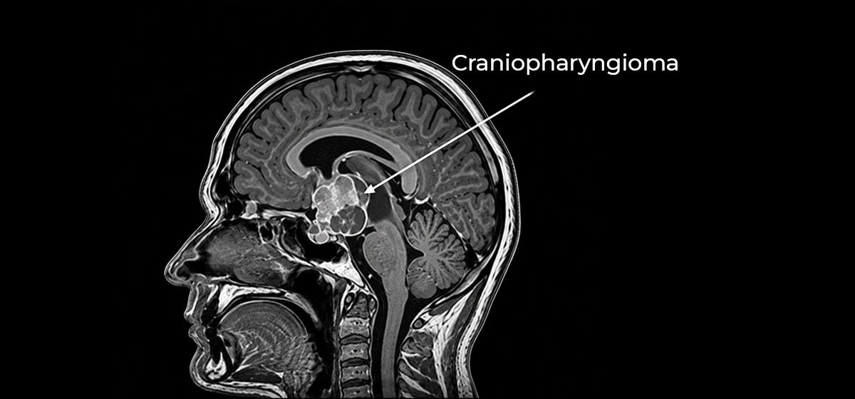
Craniopharyngioma
A skull base tumor that develops near important structures involved in vision, hormone regulation and brain function.
Learn More
Focused evaluation and treatment planning for lesions and cystic conditions involving the skull, skull base and nearby neurological structures. Care is guided by detailed clinical assessment, appropriate imaging and individualized treatment planning, with attention to the location, size and relationship of each condition to important surrounding structures.
A skull base tumor that develops near important structures involved in vision, hormone regulation and brain function.
Learn More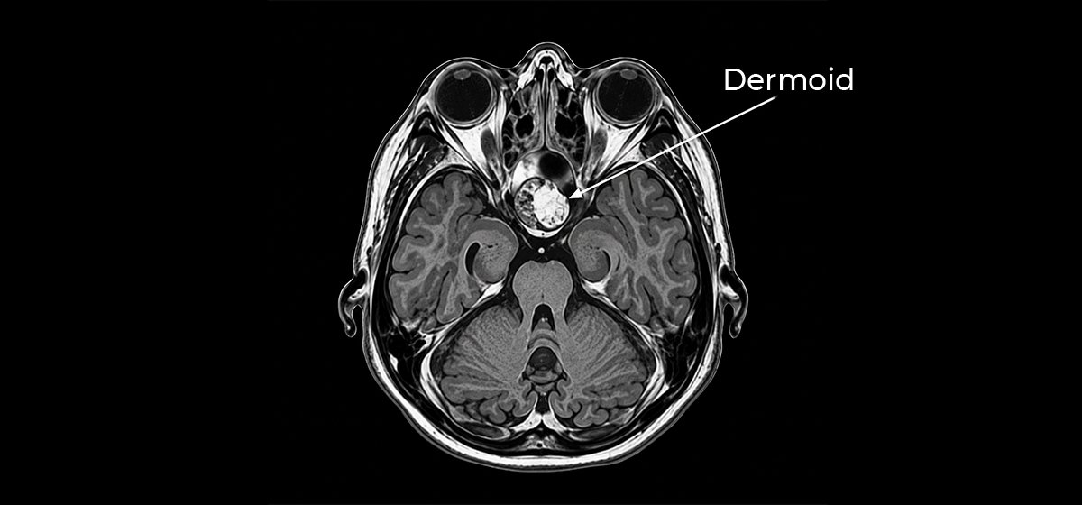
A developmental cyst that may occur around the skull base and can affect nearby structures depending on its location.
Learn More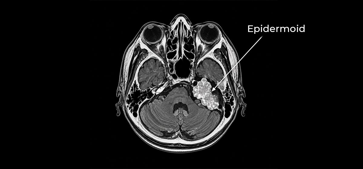
A slow-growing cystic lesion that may develop near delicate cranial nerves and other structures at the skull base.
Learn More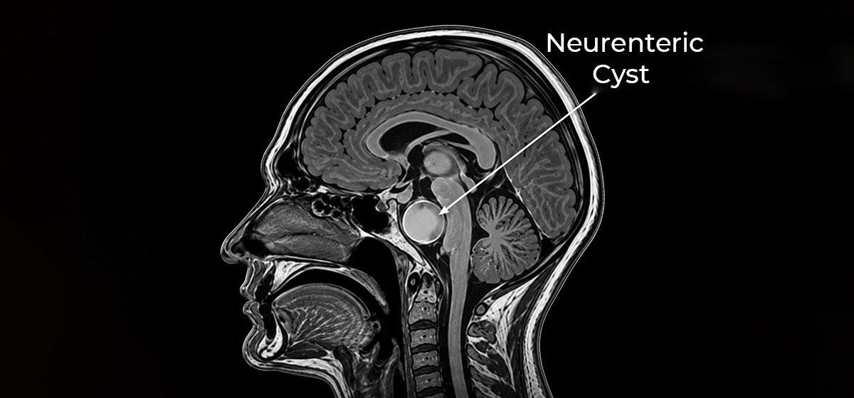
A rare developmental cyst that may occur near the brainstem, spinal cord or skull base and affect nearby structures.
Learn More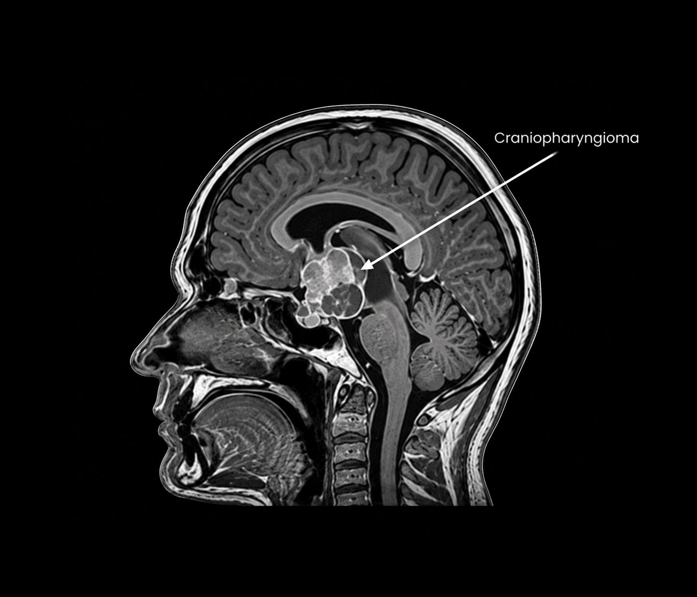
Craniopharyngioma is a tumor that develops in the region around the pituitary gland and nearby skull base structures. Because of its location, it may affect the visual pathways, pituitary function, hypothalamus and other nearby neurological structures. Symptoms can vary depending on the size and direction of growth of the lesion.
Evaluation generally includes neurological and visual assessment together with appropriate brain imaging and, when required, hormonal evaluation. Treatment planning depends on the lesion's size, location, symptoms and relationship with surrounding structures. Surgical and other treatment approaches may be considered according to the individual clinical situation.
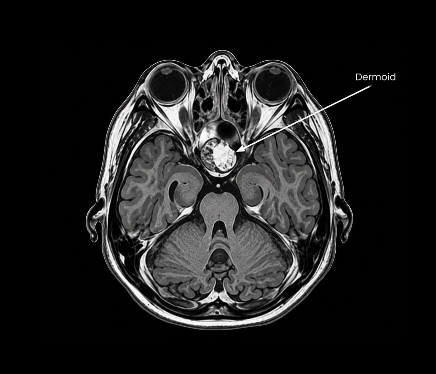
Dermoid cysts are developmental lesions that can contain tissues derived from more than one embryonic cell layer. Intracranial dermoid cysts are uncommon and may occur in locations near the skull base or other central nervous system structures. Their symptoms depend on their size, location and relationship with nearby tissues.
Evaluation typically involves clinical and neurological assessment together with appropriate CT or MRI imaging to define the lesion and its anatomical relationships. Management is individualized according to symptoms, imaging findings, lesion characteristics and its proximity to important neurological structures. Neurosurgical evaluation may be considered when clinically appropriate.
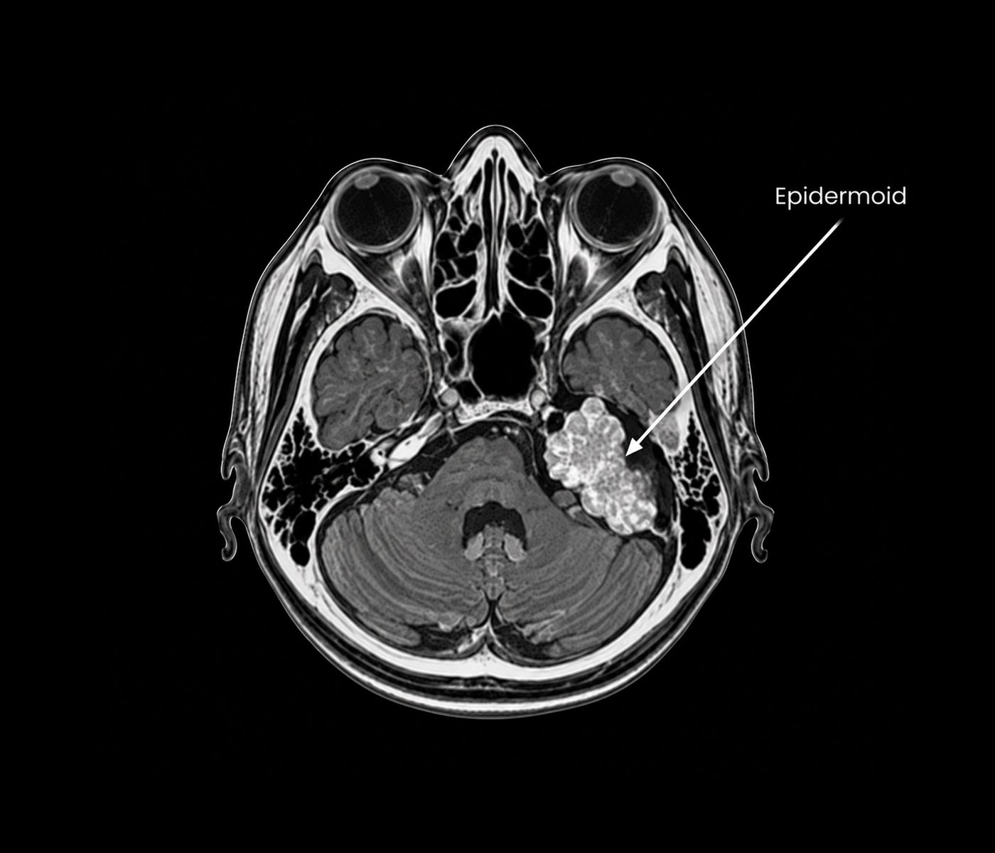
Epidermoid cysts are slow-growing lesions that develop from ectodermal tissue and may occur within the skull base or other areas of the brain. They can grow around important cranial nerves, blood vessels and other structures, and symptoms may develop gradually as the lesion affects nearby neurological anatomy.
Assessment includes neurological examination and appropriate MRI imaging to characterize the lesion and define its relationship with surrounding structures. Management depends on symptoms, lesion size and location, imaging characteristics and the degree of involvement of nearby nerves or vessels. Neurosurgical treatment may be considered when clinically indicated.
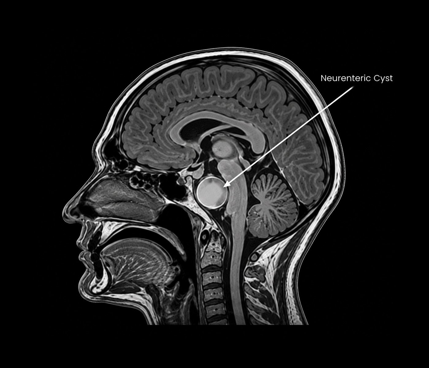
Neurenteric cysts are uncommon developmental lesions that arise from abnormalities occurring during early development. They are often located near the brainstem or spinal cord and may occasionally occur in the cranial or skull base region. Symptoms depend on the exact location and the structures affected by the cyst.
Evaluation generally includes neurological assessment and appropriate MRI imaging to determine the cyst's location, size and relationship with the brainstem, spinal cord or other nearby structures. Management is individualized based on symptoms, imaging findings and the anatomical accessibility of the lesion, with neurosurgical assessment considered when appropriate.