INTRINSIC TUMOR
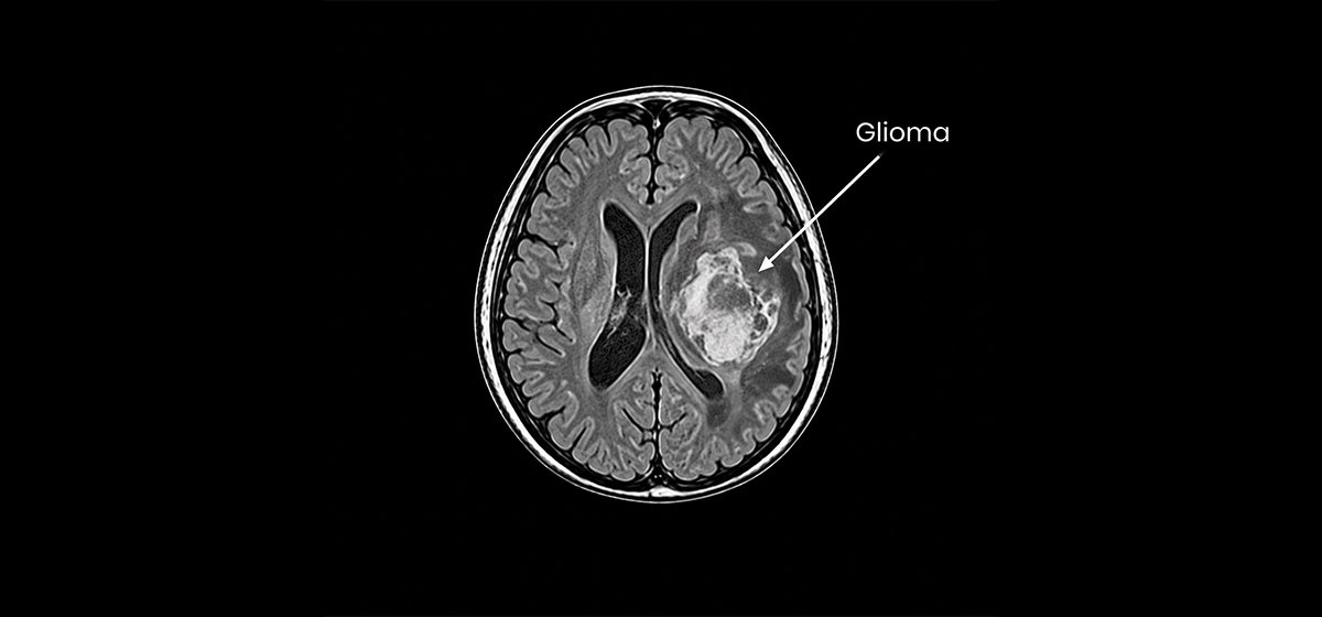
Glioma
Tumors originating within glial cells of the brain or spinal cord that require careful evaluation and planning.
Learn More
Focused evaluation and treatment planning for tumors originating within the brain and central nervous system, including gliomas, pineal tumors, ependymomas and metastatic brain tumors. Care is guided by detailed neurological assessment, appropriate imaging, tumor characteristics and individualized treatment planning.
Tumors originating within glial cells of the brain or spinal cord that require careful evaluation and planning.
Learn More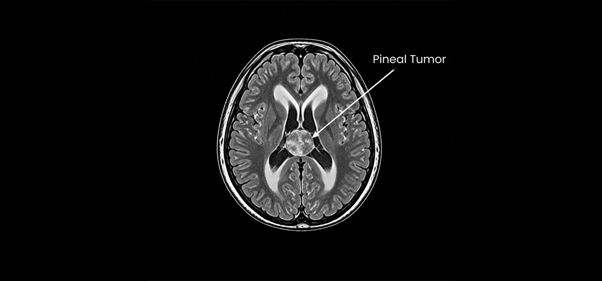
Abnormal cell growths in the pineal gland region that may affect nearby structures and cerebrospinal fluid pathways.
Learn More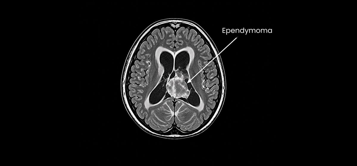
Tumors arising from cells lining the ventricular pathways that may influence cerebrospinal fluid circulation.
Learn More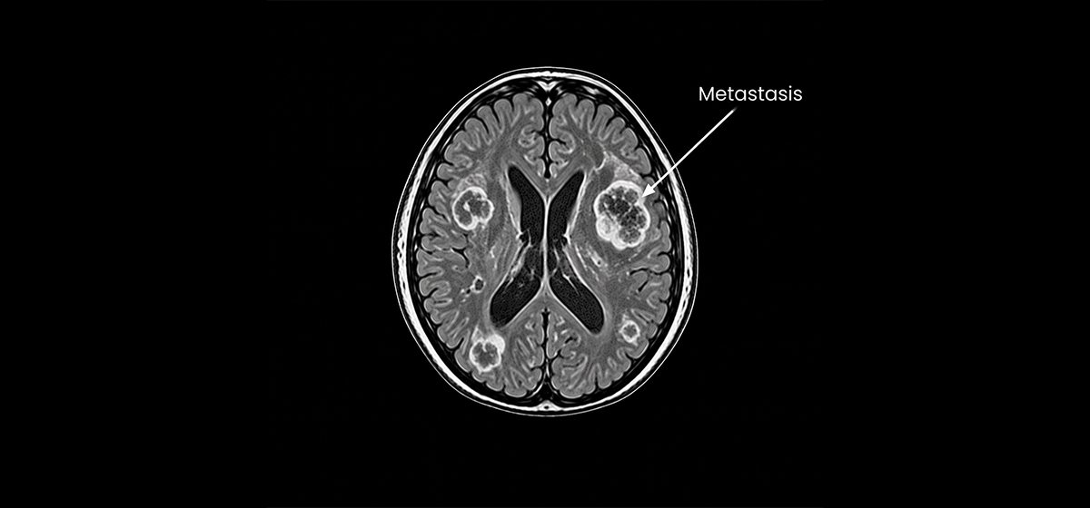
Secondary tumors that spread to the brain from cancer originating elsewhere in the body.
Learn More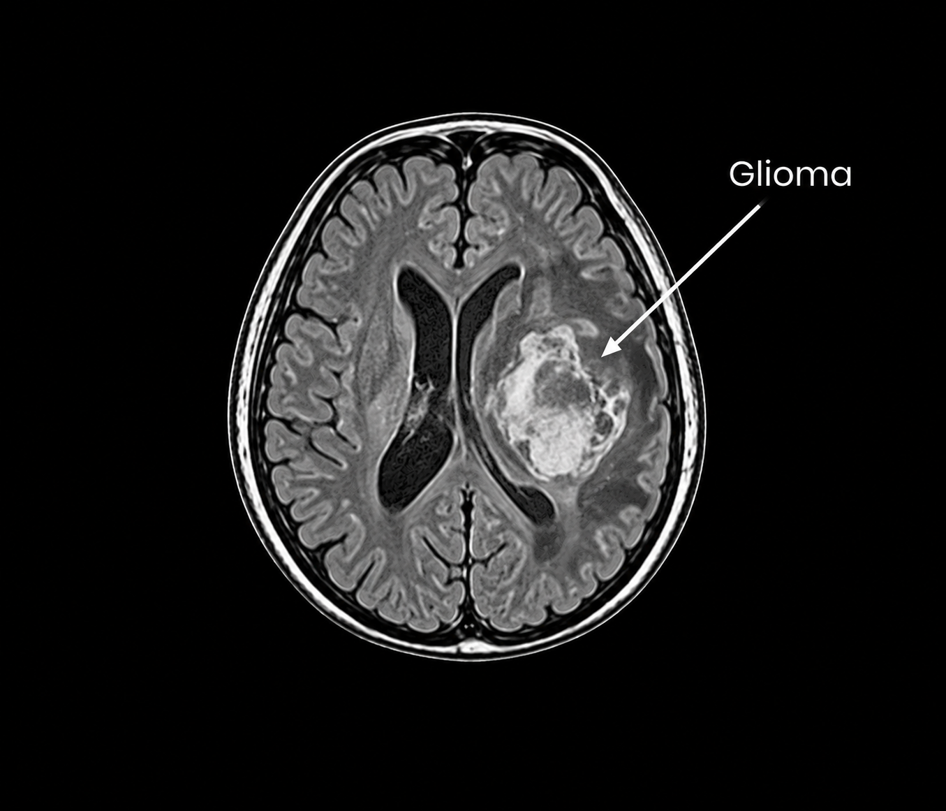
Gliomas are tumors that originate from glial cells within the brain or spinal cord. They can occur in different areas of the central nervous system and may affect important neurological functions depending on their location, size and biological characteristics. Symptoms may include headaches, seizures, weakness, changes in speech, vision, memory or coordination.
Evaluation involves detailed neurological assessment and appropriate brain or spine imaging, with further diagnostic assessment when required. Treatment planning depends on tumor location, imaging characteristics, neurological findings and the individual clinical situation. When appropriate, neurosurgical treatment may be considered as part of an individualized multidisciplinary approach.
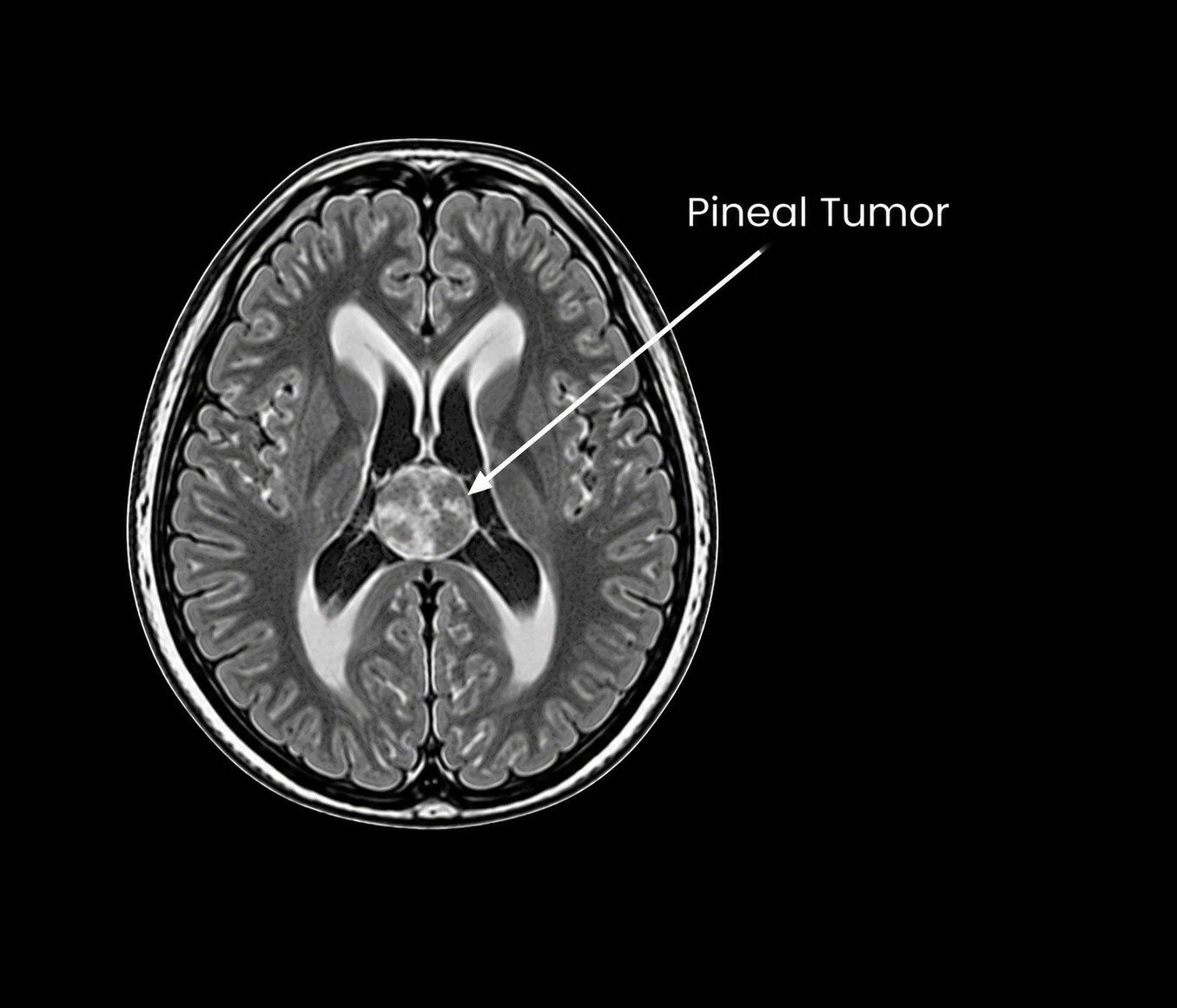
Pineal tumors are abnormal growths that develop in or around the pineal gland, a small structure located deep within the brain. Because of their position near important structures and cerebrospinal fluid pathways, these tumors may produce symptoms such as headache, nausea, visual changes or other neurological disturbances.
Evaluation typically involves detailed neurological assessment and appropriate brain imaging to understand the location, size and relationship of the lesion to surrounding structures. Treatment planning depends on the imaging findings, clinical presentation and suspected tumor type. Neurosurgical intervention may be considered when clinically appropriate as part of individualized treatment planning.
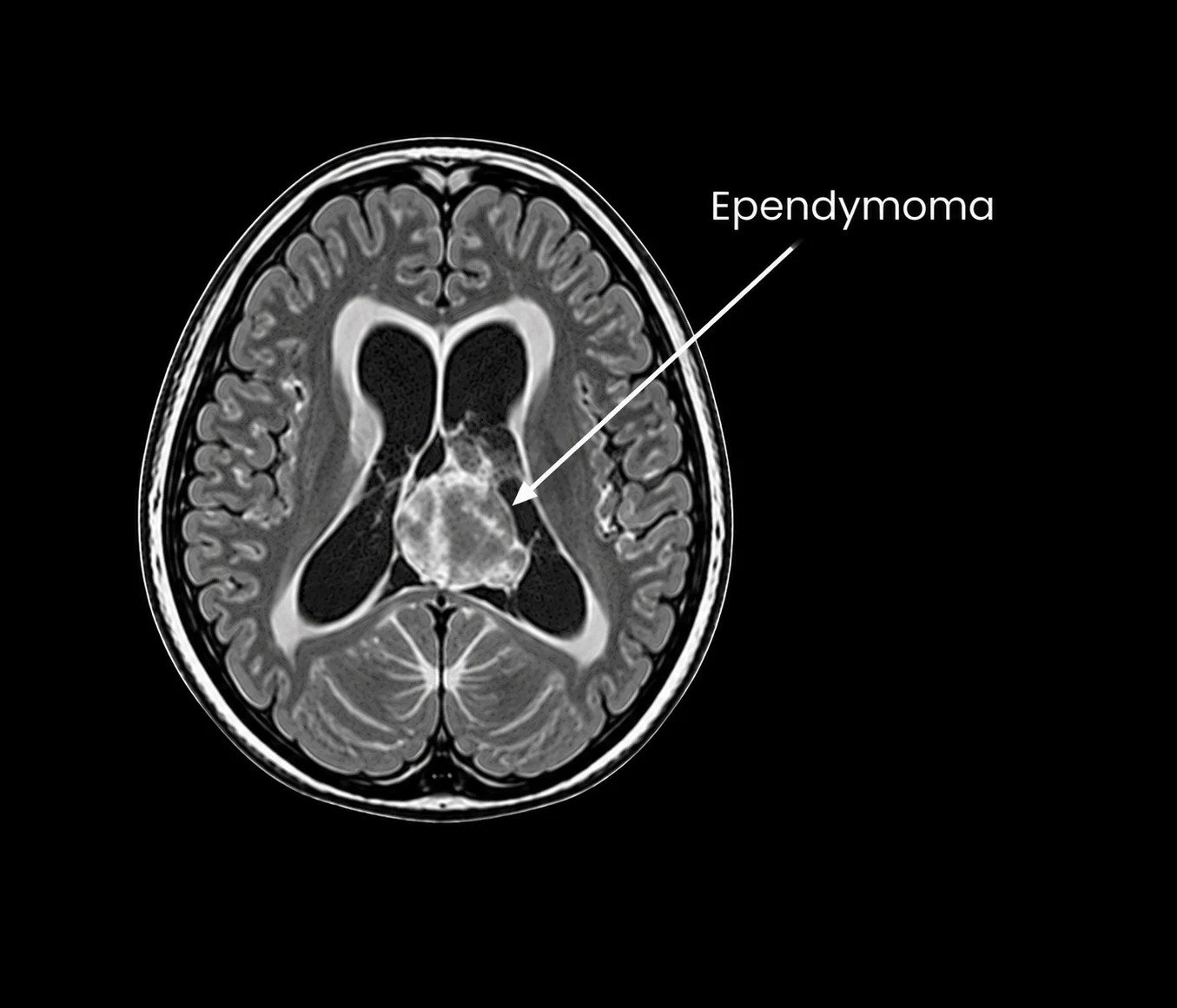
Ependymomas are tumors that arise from cells lining the ventricular pathways of the brain or the central canal of the spinal cord. When located within or near the ventricular system, they may interfere with normal cerebrospinal fluid circulation and can produce symptoms related to increased pressure or neurological dysfunction.
Assessment involves neurological examination and appropriate imaging to determine the location, size and relationship of the tumor to surrounding structures. Treatment planning is individualized according to the tumor characteristics, neurological findings and effects on cerebrospinal fluid circulation. Neurosurgical management may be considered when appropriate.
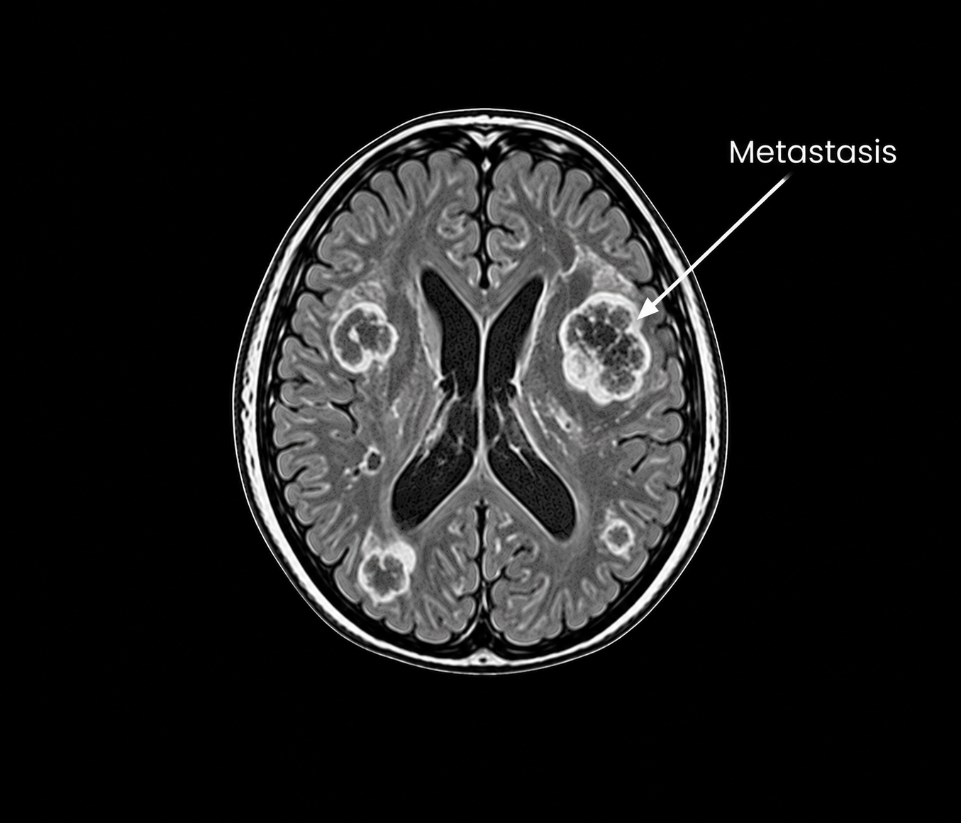
Brain metastases are secondary tumors that develop when cancer cells spread to the brain from a primary tumor elsewhere in the body. Their presentation can vary depending on the number, size and location of the lesions, with symptoms potentially including headaches, seizures, weakness, changes in speech, vision or coordination.
Evaluation involves neurological assessment and appropriate brain imaging to determine the number, location and effects of the lesions. Treatment planning depends on the underlying cancer, neurological findings, imaging characteristics and overall clinical circumstances. When appropriate, neurosurgical treatment may be considered alongside other oncological treatment approaches.