MENINGEAL TUMOR
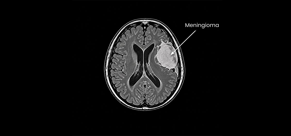
Meningioma
Evaluation of tumors arising from the protective coverings of the brain and spinal cord, with treatment planning based on location, size and clinical features.
Learn More
Focused evaluation and treatment planning for tumors that arise from structures surrounding the brain or from nearby tissues. Care is guided by neurological and endocrine assessment, appropriate imaging and individualized treatment planning, with attention to the tumor's location, size, symptoms and relationship to important neural structures.
Evaluation of tumors arising from the protective coverings of the brain and spinal cord, with treatment planning based on location, size and clinical features.
Learn More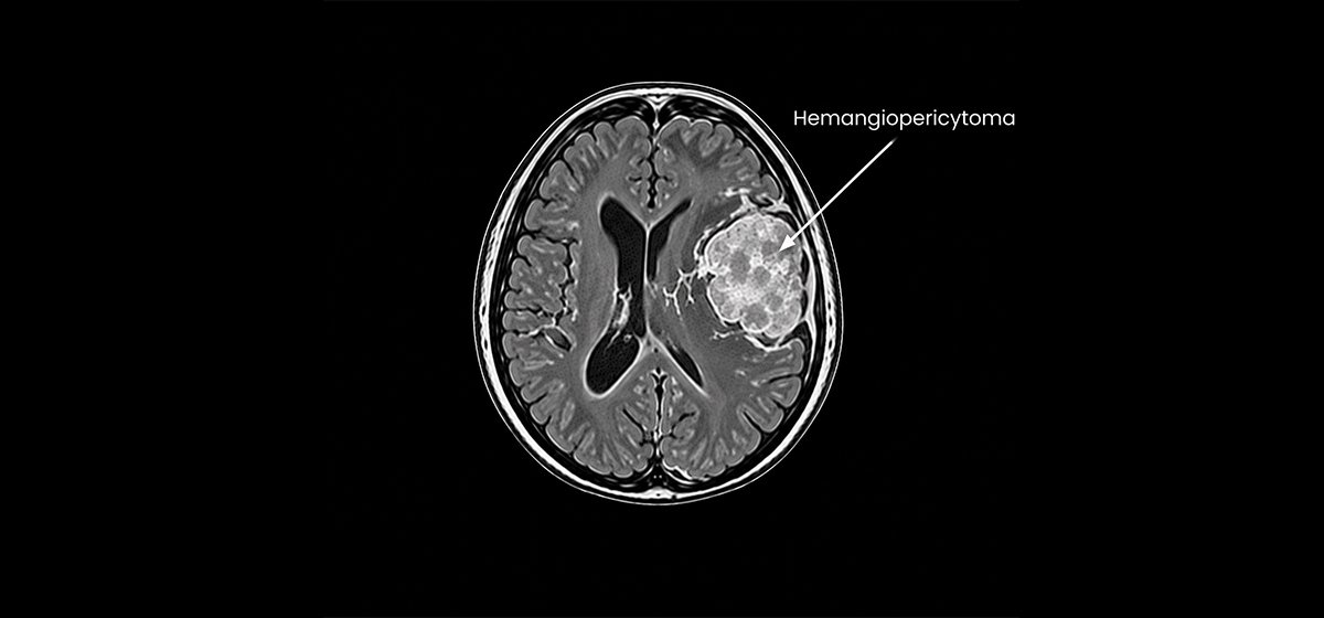
Assessment of a rare vascular tumor that may arise from the coverings of the brain and requires careful imaging and individualized neurosurgical planning.
Learn More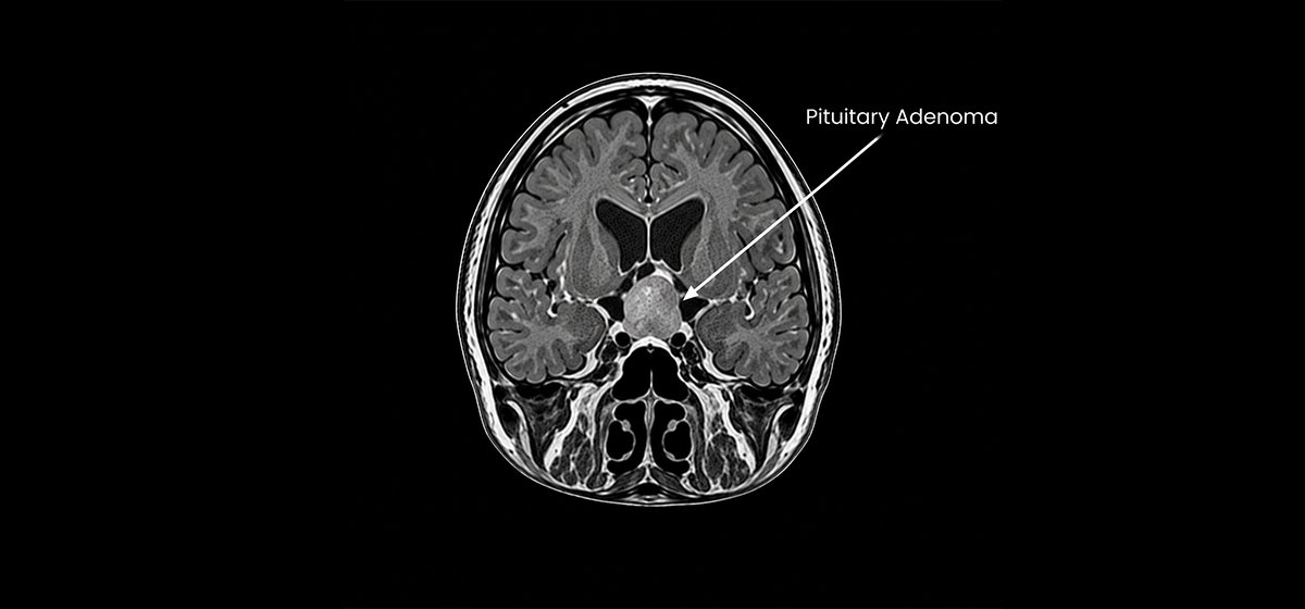
Evaluation of tumors arising from the pituitary gland, including assessment of hormonal effects, vision and the relationship of the tumor to nearby structures.
Learn More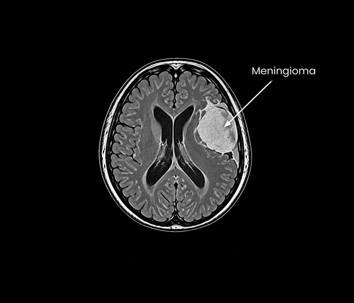
Meningiomas are tumors that arise from the meninges, the protective layers surrounding the brain and spinal cord. Many meningiomas grow slowly and may remain without symptoms for a period of time. When they affect nearby brain tissue, nerves or blood vessels, symptoms can include headache, seizures, weakness, changes in vision or other neurological difficulties depending on their location.
Evaluation involves neurological assessment and appropriate brain imaging, usually with MRI when clinically indicated, to understand the tumor's size, location and relationship with surrounding structures. Management depends on symptoms, tumor characteristics, growth pattern and overall clinical circumstances. Observation, surgical treatment or other specialist-directed approaches may be considered according to the individual case.
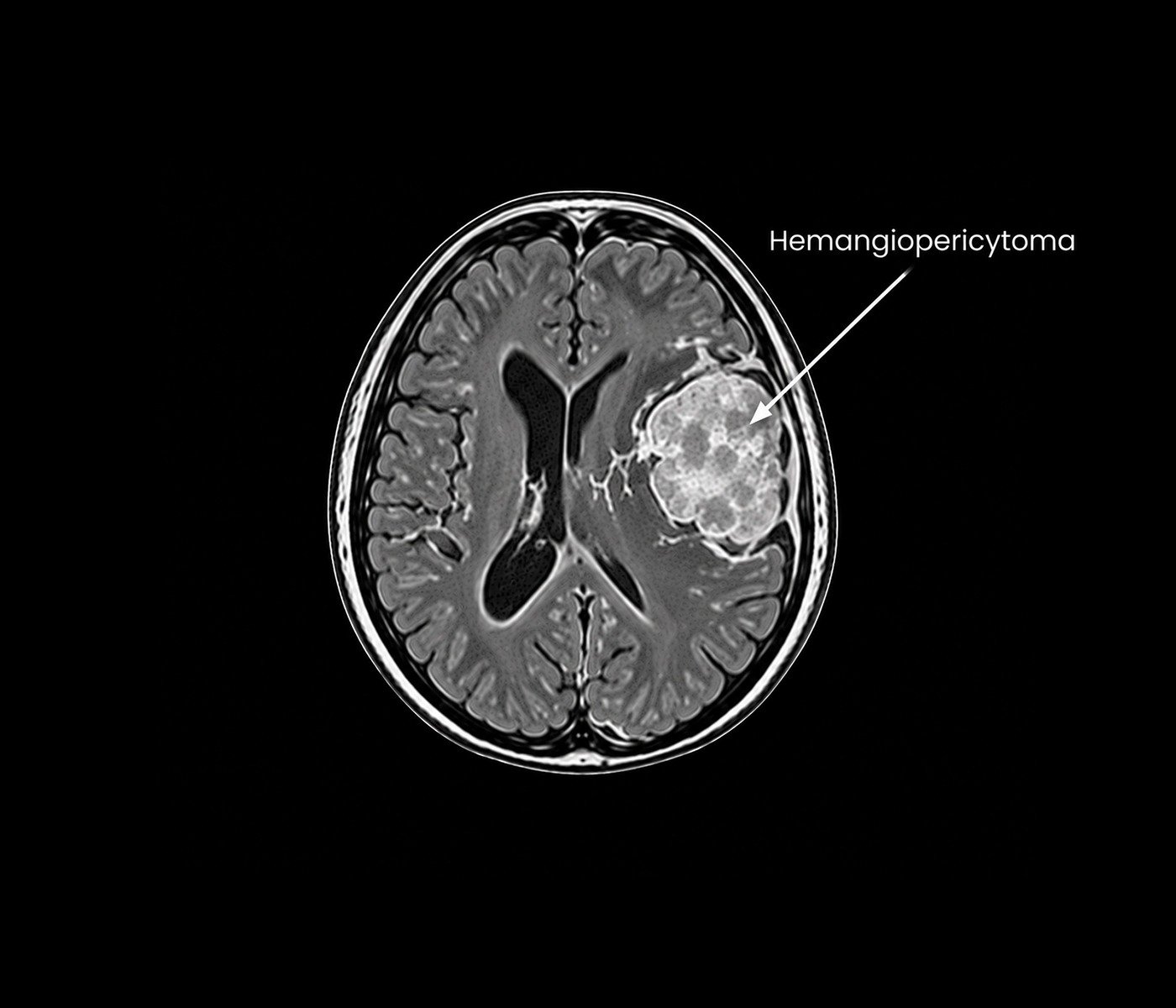
Hemangiopericytoma is a rare vascular tumor that can arise from tissues surrounding the brain. These tumors may occur in close relationship to important blood vessels, nerves and other neurological structures. Symptoms vary according to their size and location and may include headache, seizures, visual changes or focal neurological difficulties.
Evaluation requires detailed imaging to define the tumor's location, vascular characteristics and relationship with nearby structures. Treatment planning is individualized and may involve neurosurgical removal when appropriate, with careful consideration of the tumor's vascular nature and surrounding anatomy. Further management is guided by the clinical and pathological findings of each case.
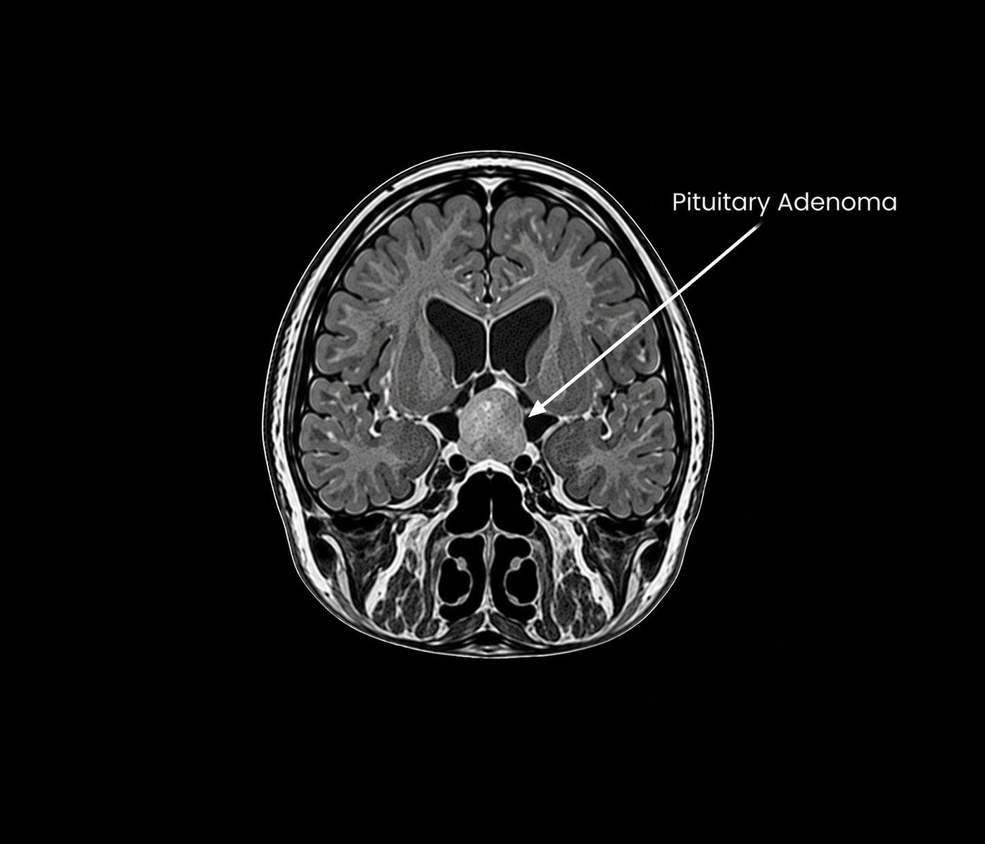
Pituitary adenomas are tumors arising from the pituitary gland, a small structure located at the base of the brain that plays an important role in regulating hormones. Some adenomas produce excess hormones, while others may affect surrounding structures as they enlarge. Symptoms can include hormonal changes, headache and visual problems, particularly when the tumor affects the nearby optic pathways.
Evaluation commonly involves brain imaging together with appropriate hormonal assessment and visual evaluation when required. Treatment planning depends on the tumor's size, hormonal activity, visual or neurological effects and overall clinical findings. Medication, observation or surgical treatment may be considered depending on the individual circumstances and specialist assessment.